Corticosteroidsreference 1,2
Modulate inflammatory pathways that are activated in DMD. Not dependent on DMD mutation type.

This site is intended for US healthcare professionals only.

To address multiple drivers of progression,
DMD is a complex disease caused by genetic mutations that result in little to no production of dystrophin, a critical structural protein in muscle fibers. This dystrophin deficiency causes muscle cell instability and disrupts key signaling pathways involved in maintaining muscle homeostasis.reference 2,3
These disruptions lead to increased histone deacetylase (HDAC) activity, which contributes to downstream pathologic processes such as chronic inflammation and dysregulated muscle repair.reference 2
Understanding these interconnected mechanisms can help support a broader view of DMD management that looks beyond dystrophin deficiency.reference 2
The dystrophin-associated protein complex (DAPC) contributes to muscle cell stability by connecting the internal cytoskeleton to the extracellular matrix and plays a key role in transmitting signals necessary to maintain muscle cell homeostasis.reference 2,3
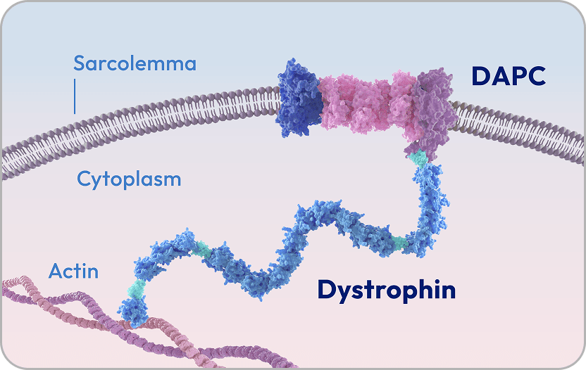
Healthy muscle with dystrophin
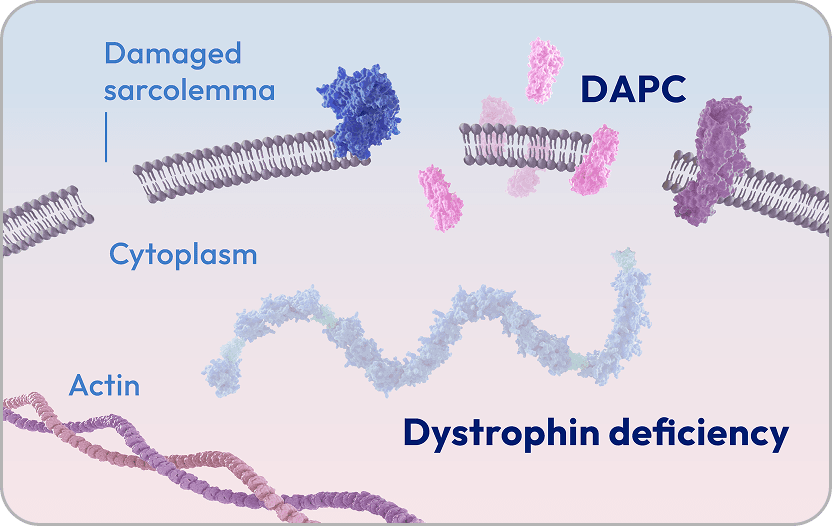
Muscle in DMD without dystrophin
Lack of dystrophin disrupts DAPC integrity, causes muscle cell instability, and leads to increased HDAC activity and disrupted cell signaling.reference 3
HDAC is an enzyme that helps regulate key processes involved in muscle integrity and repair.reference 3,6 HDAC acts by deacetylating proteins in both the nucleus and cytoplasm, supporting gene transcription, protein function, and signaling pathways involved in muscle homeostasis.reference 3,6-8
In the nucleus, HDAC plays a role in muscle repair by acting on (modifying) histone proteins and regulating gene transcription.
In the cytoplasm, HDAC influences muscle integrity and stability by acting on (modifying) nonhistone proteins.
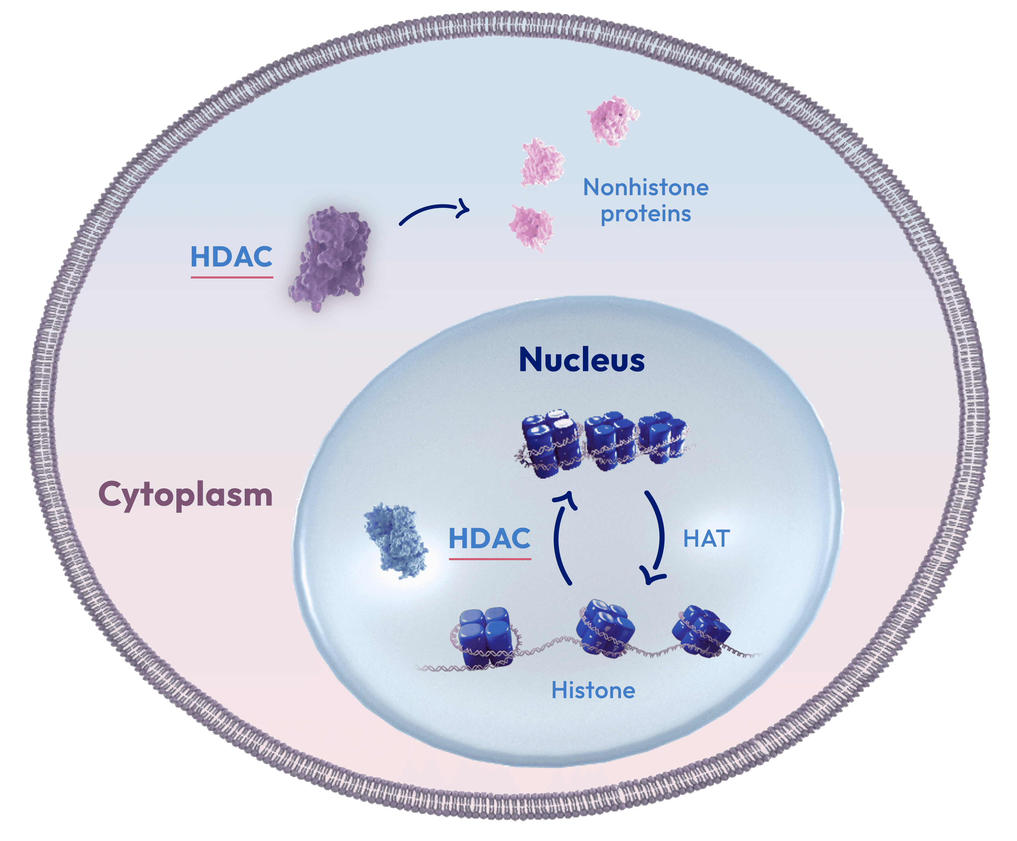
HAT, histone acetyltransferase.
Altogether, overactive HDAC contributes to several key hallmarks of DMD, includingreference 2,3,12:

Persistent activation of chronic inflammatory pathways

Dysregulated muscle repair due to disrupted gene transcription and decreased stem cell differentiation

Fibrogenesis and adipogenesis

Progressive muscular atrophy
Persistent activation of chronic inflammatory pathways
HDAC overactivity is a key driver of DMD and plays a causative role in disease progression.reference 2,3,12
Given the multifactorial nature of DMD, management may require approaches that address more than one disease mechanism.reference 1,2
Stay informed about treatment approaches in DMD.
Sign up for updatesDMD is a multifactorial disease that may require a comprehensive treatment approachreference 1,2
Because DMD involves multiple pathologic processes, no single treatment approach addresses all aspects of the disease.reference 1,2
A comprehensive management strategy may include:
Together, these approaches may help to more fully address the complex and evolving challenges of DMD.
Different treatments are thought to act on different specific aspects of DMD pathophysiology:
Modulate inflammatory pathways that are activated in DMD. Not dependent on DMD mutation type.
Different treatments are thought to act on different specific aspects of DMD pathophysiology:
Modulate inflammatory pathways that are activated in DMD. Not dependent on DMD mutation type.
Target specific exon mutations to enable production of shortened forms of dystrophin. These types of therapies are dependent on DMD mutation type.

Delivers a gene designed to enable production of microdystrophin, a shortened form of the dystrophin protein. Suitable for many DMD mutation types.
Designed to reduce HDAC overactivity associated with DMD. Not dependent on DMD mutation type.

Being a multisystemic disease, DMD requires a multidisciplinary care approach. Although the composition of the care team may vary and change over time, specialists across disciplines can help patients address the complex and evolving challenges of DMD.reference 13,16
Supportive therapy in DMD, such as physical therapy and occupational therapy, can also play an important role in helping to preserve function.reference 13
A closer look at how each MDT member informs the patient care ecosystemreference 16:





The complexity of DMD supports a multimodal treatment strategy and multidisciplinary care approach that address multiple disease mechanisms as well as patient needs.reference 2,13,16
Complete the form below to receive additional content and resources from ITF Therapeutics—including information about a treatment option for your patients.
*Required field.
References: 1. Duan D, Goemans N, Takeda S, Mercuri E, Aartsma-Rus A. Duchenne muscular dystrophy. Nat Rev Dis Primers. 2021;7(1):13. 2. Montagna C, Maiani E, Pieroni L, Consalvi S. Duchenne muscular dystrophy: integrating current clinical practice with future therapeutic and diagnostic horizons. Int J Mol Sci. 2025;26(14):6742. 3. Consalvi S, Saccone V, Giordani L, Minetti G, Mozzetta C, Puri PL. Histone deacetylase inhibitors in the treatment of muscular dystrophies: epigenetic drugs for genetic diseases. Mol Med. 2011;17(5-6):457-465. 4. Kodippili K, Rudnicki MA. Satellite cell contribution to disease pathology in Duchenne muscular dystrophy. Front Physiol. 2023;14:1180980. 5. Dowling P, Swandulla D, Ohlendieck K. Cellular pathogenesis of Duchenne muscular dystrophy: progressive myofibre degeneration, chronic inflammation, reactive myofibrosis and satellite cell dysfunction. Eur J Transl Myol. 2023;33(4):11856. 6. Seto E, Yoshida M. Erasers of histone acetylation: the histone deacetylase enzymes. Cold Spring Harb Perspect Biol. 2014;6(4):a018713. 7. Yang C, Croteau S, Hardy P. Histone deacetylase (HDAC) 9: versatile biological functions and emerging roles in human cancer. Cell Oncol (Dordr). 2021;44(5):997-1017. 8. Tian H, Liu S, Ren J, et al. Role of histone deacetylases in skeletal muscle physiology and systemic energy homeostasis: implications for metabolic diseases and therapy. Front Physiol. 2020;11:949. 9. Rugowska A, Starosta A, Konieczny P. Epigenetic modifications in muscle regeneration and progression of Duchenne muscular dystrophy. Clin Epigenetics. 2021;13(1):13. 10. Colussi C, Mozzetta C, Gurtner A, et al. HDAC2 blockade by nitric oxide and histone deacetylase inhibitors reveals a common target in Duchenne muscular dystrophy treatment. Proc Natl Acad Sci USA. 2008;105(49):19183-19187. 11. Marrone AK, Shcherbata HR. Dystrophin orchestrates the epigenetic profile of muscle cells via miRNAs. Front Genet. 2011;2:64. 12. Huang Z, Hu L, Liu Z, Wang S. The functions and regulatory mechanisms of histone modifications in skeletal muscle development and disease. Int J Mol Sci. 2025;26(8):3644. 13. Parent Project Muscular Dystrophy. Assembling a care team. Parent Project Muscular Dystrophy. Accessed July 9, 2026. https://www.parentprojectmd.org/care/for-families/assembling-a-care-team. 14. Happi Mbakam C, Lamothe G, Tremblay JP. Therapeutic strategies for dystrophin replacement in Duchenne muscular dystrophy. Front Med (Lausanne). 2022;9:859930. 15. Chwalenia K, Feng VY, Hemmer N, et al. AAV microdystrophin gene replacement therapy for Duchenne muscular dystrophy: progress and prospects. Gene Ther. 2025;32(5):447-461. 16. Birnkrant DJ, Bushby K, Bann CM, et al. Diagnosis and management of Duchenne muscular dystrophy, part 1: diagnosis, and neuromuscular, rehabilitation, endocrine, and gastrointestinal and nutritional management. Lancet Neurol. 2018;17(3):251-267.